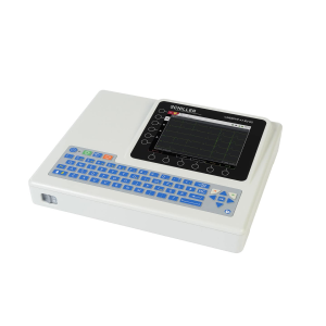
Intelligent Automatic functions for smooth workflow
- Auto Optimizer: Automatically adjusts the gain and baseline position and velocity range with just one button
- Cardiac Features: automation tools enable faster and smoother cardiovascular examination, building on data acquired by our premium systems
- Mobility that meets autonomy – allowing to scan approx. 1 hour in battery mode for smooth use in emergency care or changing rooms
- Power cord hook, handles and folding mechanism, allowing to move the system safely
Superb Imaging, powered by top-performing imaging technologies
Top-performing imaging technologies have been mitigated for enhanced diagnostic confidence and precision, namely:
- High definition dynamic Tissue Harmonic Imaging (HdTHI) resulting in superb spatial resolution and penetration
- High Resolution Imaging (HI REZ) to emphasize tissue structures and enhance contrast resolution for greater clarity
- Compound Imaging for clear visualization of tissue boundaries, high contrast resolution and speckle reduction to allow clearer observation of lesions
Simple to use applications, enhancing precision in a wide user range
ARIETTA 65 incorporates advanced tools for diverse clinical use and detailed evaluation:
- Real-time Tissue Elastography(RTE)
- Assesses tissue strain in real time and differences in tissue stiffness as a color map – useful in a wide variety of clinical fields such as breast, thyroid gland and urinary structures.
- Estimates liver fibrosis staging in patients with hepatitis C (LF Index) with the abdominal convex transducer
- Supports time-saving automation features to improve objectivity, reproducibility and productivity
- Contrast Harmonic Imaging (CHI) – provides homogeneous enhancement throughout the field of view to enhance diagnostic capability.
- Panoramic View – generates a single, elongated image by moving the probe across the target organ, thus allowing a larger field of view and enhanced diagnostic precision.
Interventional support tools
- Needle Emphasis – automatically adjusts the deflection angle of beams and images to enhance needle visibility and assist in safe and accurate punctures.
- Marking Assist to support marking on the body surface before surgical operation.
Automatic tools for vascular and cardiac
Auto IMT
- Automatically measures the max and mean values of Intima-Media Thickness (IMT) for reproducible and accurate follow up of vascular disease
Dual Gate Doppler
- Collects Doppler waveforms from two locations during the same heart beat
- Enables faster and more accurate measurement of LV diastolic performance indicators
- Useful in diagnosis of fetal arrhythmia and Carotid stenosis
2D Tissue Tracking (2DTT)
- Speckle tracking technique that quantifies the movement of the entire left ventricle or the local movement of the myocardium resulting in dynamic analysis of myocardium.